About the webinar:
Next-generation targeted anticancer therapies demand advanced preclinical models that recapitulate the tumor microenvironment. Research shows that advanced orthotopic patient-derived xenograft (PDX) models:
- Accurately predict drug response, drug resistance and metastatic proliferation
- Reliably mirror the behavior of cancer in their human counterparts, at an accelerated pace — making them clinically relevant patient avatars for evaluating cancer therapies
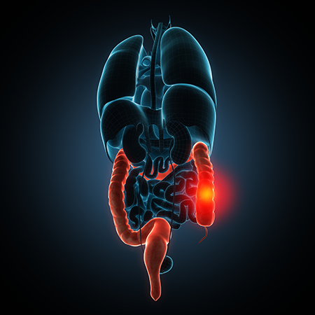
Developing orthotopic PDX models involves surgically implanting a very small sample of human tumor tissue in the corresponding anatomic location (colon, brain, pancreas, liver, lung, etc.) of specialized immunodeficient NOG mice, effectively creating a living model of that unique cancer. Certis scientists have spent years perfecting the art of orthotopic PDX engraftment, using novel surgical techniques and sophisticated animal husbandry protocols to optimize cohort viability.
Watch this on-demand webinar as Certis Oncology Chief Scientific Officer Jonathan Nakashima discusses how to move the most promising drug candidates forward to the clinic efficiently and effectively with advanced orthotopic PDX models of cancer.
Attend the webinar to:
- Discover how the tumor microenvironment factors into selecting optimal cancer model(s) for your preclinical in vivo pharmacology study
- Understand how orthotopic PDX models combined with live in situ imaging (like mouse MRI) can accurately predict drug response, drug resistance and metastatic proliferation
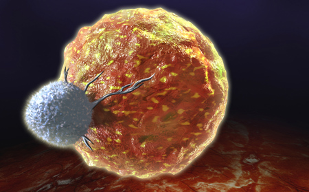
- Uncover how the need for clinically relevant solid tumor models for the translation of T cell-based therapies into the clinic
- Learn how orthotopic PDX models can be used to validate artificial intelligence predictions in drug discovery and development
- Gain knowledge on personalized orthotopic PDX of an individual’s cancer can be used as a functional testing platform







.jpg)

.jpg)
.jpg)
.jpg)
.jpg)


.jpg)


.jpg)
.jpg)


.jpg)


.jpg)

.jpg)


