New Rab29 Overexpression Mouse Model for Parkinson's Disease

Terina Martinez, PhD
Tuesday, January 14th, 2020
Parkinson's disease (PD) is the second most common neurodegenerative disease (after Alzheimer's disease) and the most common movement disorder. The worldwide incidence of PD has more than doubled since 1990 to an estimated 6.1 million PD patients globally in 20161. In the United States in 2017, the estimated total economic burden of PD was $51.9 billion (The Lewin Group, Inc., Final Report commissioned by and submitted to The Michael J. Fox Foundation for Parkinson's Research).

PD has a complex etiology, underscored by multifaceted interactions between genetics and environment on the background of aging. Among the more than a dozen genes linked to Mendelian inherited forms of PD, mutations in the gene encoding the protein leucine-rich repeat kinase 2 (LRRK2) are associated with autosomal dominant risk for PD2. Moreover, RAB29 is one of five genes identified within the PARK16 locus that has been linked to human PD2,3.
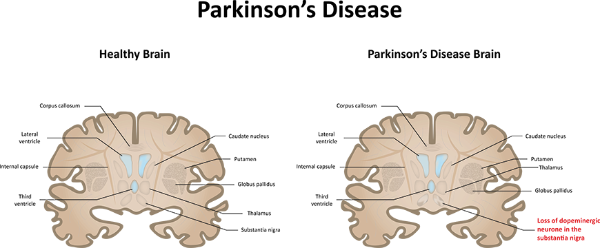
Comparison between healthy brain and brain inflicted with Parkinson's Disease
Often, a lack of ideal research tools and models are limiting factors in testing new scientific concepts. When scientists must make their own research tools, it inefficiently expends time and resources and constrains replicability, quality control, and wide access to the tools. Through its research tools program, The Michael J. Fox Foundation for Parkinson's Research (MJFF) addresses this by partnering with field-wide experts to identify what tools are missing from the toolbox and working with qualified vendors to produce and distribute the tools without restrictions to access.

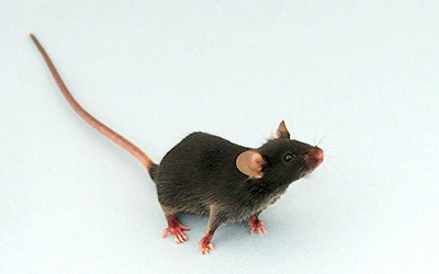
In its continued efforts to facilitate access to improved preclinical models for parkinsonism, MJFF partnered with Taconic Biosciences to generate a new genetically engineered mouse model for PD, culminating in the recent launch of the Rab29 Overexpression Mouse model (Taconic Model #16552).

Taconic Launches a Rab29 Overexpression Mouse Model for Parkinson's Disease

The ideal preclinical model for Parkinson's disease (PD) would meet the following criteria:

  • Displays insidious multi-domain symptomology emergence (both within the central nervous system and peripheral compartments) exacerbated by aging
  • Presents a progressive pathophysiology
  • Has the ability to recapitulate the stereotyped motor deficits of the human condition, in a manner that is reversible by exogenous dopamine
  • Involves pathology within the dopaminergic neuron cell bodies and neural terminals of the nigrostriatal system as well as within non-nigral neural nuclei
Essentially, satisfying the above criteria would recapitulate with reasonable fidelity the symptomology and pathological effects observed in human PD cases.

LRRK2 in familial and idiopathic PD

Accounting for approximately 5% of familial PD cases, LRRK2 represents one of the most commonly mutated genes linked to heritable PD and is also observed to be mutated in approximately 1% of sporadic PD patients4. Due to its linkage with genetic cases of PD as well as its association with sporadic PD and combined with its ubiquitous expression and putative kinase function, LRRK2 has emerged as an attractive candidate for disease-modifying therapeutic development for PD5.

Structure and Function of LRRK2

LRRK2 is a large, multi-domain protein kinase consisting of an armadillo repeat domain, an akyrin domain, leucine-rich repeats, a ROC-type GTPase domain that closely resembles a Rab GTPase and is associated with a COR domain, a serine/threonine protein kinase domain, and a WD40 repeat-containing domain6. The most common pathogenic mutation in LRRK2 — G2019S — lies within the catalytic domain and increases LRRK2 kinase activity7-9.

Taconic offers two MJFF-sponsored LRRK2 G2019S models: a LRRK2 G2019S KI Mouse model with targeted insertion of the mouse LRRK2 gene harboring the G2019S mutation and a human LRRK2 G2019S Rat model, with random insertion of the human LRRK2 gene into the Sprague Dawley® rat.

Functionally, LRRK2 has been implicated in numerous pathways in PD, including inflammation and immune system dysfunction10,11, aberrant autophagosome, endosome, and lysosome trafficking/function12, and synapse development, maturation, and function13, among others.

Identification of Rab Proteins as LRRK2 Substrates

While the toxic gain of function by increased LRRK2 kinase activity downstream of the G2019S mutation was appreciated for over a decade, bona fide, endogenous substrates of LRRK2's kinase activity long-eluded the PD research community. In the last couple of years however, phosphoproteomic analyses revealed a subset of Rab GTPases regulated by LRRK214. Ten Rab proteins were further shown to be endogenous substrates of LRRK2 and/or regulators of LRRK2 signaling, including Rab8a, Rab10, Rab12, Rab29, and Rab3514,15. Notably, Rab29 is also known as Rab7L1. More information on Rab29 nomenclature can be found at Rab29 GeneCard, UniProt, and Gene - NCBI.

Physiological and Pathophysiological Roles of Rab29

Rab GTPases are master regulators of membrane trafficking. In the majority of cases reported to date, Rab GTPase phosphorylation is predicted to impede Rab protein function; however, the discovery of LRRK2 as a protein exhibiting preferential binding to phosphorylated Rabs and subsequent increased LRRK2 activity suggests that LRRK2-mediated pathology may be attributed to more complex Rab protein interactions than previously appreciated16.

Since its discovery and validation as a substrate of LRRK2 kinase activity, Rab29 has emerged as a master regulator of LRRK2 signaling and has been implicated in the following cellular functions/pathways:

  • Primary ciliogenesis17
  • Membrane trafficking18
  • Retromer complex assembly/function19
  • Axonal morphology/neurite outgrowth18,20
  • Golgi complex recruitment/modification of trans-Golgi morphology21,22
  • Lysosomal homeostasis20,23
  • Centrisome defects24

Implications for Rab29-related LRRK2 Signaling in PD

Mutations that prevent LRRK2 from interacting with either Rab29 or GTP robustly inhibit LRRK2 phosphorylation at a number of important sites (including Ser910, Ser935, Ser955, and Ser97315), many of which are markers of toxic LRRK2 function25,26 or are associated with pathogenic LRRK2 mutations27.

Moreover, by nature of its regulation of LRRK2 activity, Rab29 is potentially implicated by association with aberrant LRRK2-related functions in PD, including defective ciliogenesis and centromere function, axonal/neurite morphology, Golgi and lysosomal homeostasis, etc., but the specific role of Rab29 in some of these LRRK2-related pathways may not yet be established in the published literature. As the discovery of the Rab29-LRRK2 axis was only recently described, many gaps in Rab29-mediated homeostasis and Parkinson's disease-associated pathogenic signaling remain to be explored. Research tools like the Rab29 Overexpression Mouse model will be instrumental in such studies.

The Rab29 Toolbox for PD Researchers

Taconic's launch of the PD-relevant Rab29 Overexpression Mouse is part of a growing collection of preclinical research tools in the Rab29 space. In addition to supporting the development of this Rab29 Overexpression Mouse now available at Taconic, MJFF — in partnership with Abcam Inc., and in collaboration with the Alessi Lab at the University of Dundee — also sponsored the generation and commercial distribution of custom-made rabbit monoclonal antibodies to detect total and phosphorylated versions of Rab proteins, including Rab29. These antibodies are available at Abcam and have been tested and validated in multiple applications (e.g., immunoprecipitation, immunoblotting, and immunocytochemistry) for detecting Rab protein expression in diverse cells and tissues28. Moreover, anti-Rab10 antibodies have been successfully deployed to detect total and phosphorylated Rab10 in human neutrophils29, tangibly demonstrating the conserved expression and function of LRRK2-relevant Rab proteins across mouse and human phylogeny and the tantalizing potential for development of relevant LRRK2-Rab biomarker assays. However, the assessment of pRab29 in human leukocytes has not yet been reported in the published literature.

Prospective Rab29 Research — Role in PD Novel Diagnostics and Therapies

Notably, LRRK2 kinase activity is aberrantly increased in dopamine neurons in postmortem human brain tissue from idiopathic PD cases30. This further supports the notion that LRRK2 kinase inhibitors and potentially inhibitors of Rab29 (due to its role in regulation of LRRK2 activity) may be useful therapeutics for idiopathic PD patients as well as PD patients harboring pathogenic LRRK2 mutations.

Collectively, LRRK2-related Rab signaling is an exciting and emerging new direction in Parkinson's disease research and therapeutic development. Early stage clinical trials testing LRRK2 kinase inhibitors in PD patients are already underway. Further understanding of Rab29-regulated LRRK2 kinase activation in clinically relevant paradigms will likely be instrumental in accelerating drug and biomarker discovery for PD; in addition to providing insight into LRRK2-mediated pathology in PD, Rab29-mediated LRRK2 signaling may also be amenable for leverage as a diagnostic biomarker or for therapeutic engagement in LRRK2 mutation-driven or idiopathic versions of PD.

Rab29 is a very new player in the PD landscape and much remains to be studied in this nacent sub-field. Taconic's Rab29 Overexpression Mouse model is a new and important tool in the toolbox for this emerging area of Parkinson's disease research.

Additional PD-relevant models available

MJFF has sponsored other Parkinson's disease-related animal models that are available at Taconic, including BAC transgenic rats expressing mutant versions of alpha-synuclein or LRRK2, rat TH-GFP reporter models, and a knock-in mouse model for human LRRK2. Taconic Biosciences has information on Parkinson's disease models within the neuroscience portfolio.

References:
1. GBD 2016 Parkinson's Disease Collaborators. Global, regional, and national burden of Parkinson's disease, 1990-2016: a systematic analysis for the Global Burden of Disease Study 2016. Lancet Neurol. 2018 Nov;17(11):939-953.
2. Simon-Sanchez, J, et al. Genome-wide association study reveals genetic risk underlying Parkinson's disease. Nat Genet. 2009 Dec;41(12):1308-12.
3. Tucci A, Nalls MA, Houlden H, Revesz T, Singelton, AB, Wood NW, Hardy J, Paisan-Ruiz C. Genetic variability at the PARK16 locus. Eur J Hum Genet. 2010 Dec;18(12):1356-9.
4. Healy DG, et al. Phenotype, genotype, and worldwide genetic penetrance of LRRK2-associated Parkinson's disease: a case-control study. Lancet Neurol. 2008 Jul;7(7):583-90.
5. Hatcher JM, Choi HG, Alessi DR, Gray NS. Small-molecule inhibitors of LRRK2. Adv Neurobiol. 2017;14:241-264.
6. Guaitoli G, Raimondi F, Filsbach BK, Gomez-Llorente Y, Deyaert E., Renzi F, Li X, Schaffner A, Jagtap PK, vonZweydorf F, Gotthardt, K, Lorimer DD, Yue Z, Burgin A, Janjic N, Sattler M, Versees W, Ueffing M, Ubarretxena-Belandia I, Kortholt A, Gloeckner CJ. Structural model of the dimeric Parkinson's protein LRRK2 reveals a compact architecture involving distant interdomain contacts. Proc Natl Acad Sci U S A. 2016 Jul 26;113(30):E4357-66.
7. Ozelius LJ, Senthil G, Saunders-Pullman R, Ohmann E, Deligtisch A, Tagliati M, Hunt AL, Klein C, Henick B, Hailpern SM, Lipton RB, Soto-Valencia J, Risch N, Bressman SB. LRRK2 G2019S as a cause of Parkinson's disease in Ashkenazi Jews. N Engl J Med. 2006 Jan 26;354(4):424-5.
8. Greggio E, Jain S, Kingsbury A, Bandopadhyay R, Lewis P, Kaganovich A, van der Brug MP, Beilina A, Blackinton J, Thomas KJ, Ahmad R, Miller DW, Kesavapany S, Singleton A, Lees A, Harvey RJ, Harvey K, Cookson MR. Kinase activity is required for the toxic effects of mutant LRRK2/dardarin. Neurobiol Dis. 2006 Aug;23(2):329-41.
9. Smith WW, Pei Z, Jiang H, Dawson VL, Dawson TM, Ross CA. Kinase activity of mutant LRRK2 mediates neuronal toxicity. Nat Neurosci. 2006 Oct;9(10):1231-3.
10. Kozina E, Sadasivan S, Jiao Y, Duo Y, Ma Z, Tan H, Kodali K, Shaw T, Peng J, Smeyne RJ. Mutant LRRK2 mediates peripheral and central immune responses leading to neurodegeneration in vivo. Brain. 2018 Jun 1;141(6):1753-1769.
11. Dzamko, NL, LRRK2 and the Immune System. Adv Neurobiol. 2017;14:123-143.
12. Roosen, DA, Cookson MR. LRRK2 at the interface of autophagosomes, endosomes, and lysosomes. Mol Neurodegener. 2016 Dec 7;11(1):73.
13. Benson DL, Matikainen-Ankney BA, Hussein A, Huntley GW. Functional and behavioral consequences of Parkinson's disease-associated LRRK2-G2019S mutation. Biochem Soc Trans. 2018 Dec 17;46(6):1697-1705.
14. Steger M, Tonelli F, Ito G, Davies P, Trost M, Vetter M, Wachter S, Lorentzen E, Duddy G, Wilson S, Baptista MA, Fiske BK, Fell MJ, Morrow JA, Reith AD, Alessi DR, Mann M. Phosphoproteomics reveals that Parkinson's disease kinase LRRK2 regulates a subset of Rab GTPases. Elife. 2016 Jan 29;5. pii: e12813.
15. Purlyte E, Dhekne HS, Sarhan AR, Gomez R, Lis P, Wightman M, Martinez TN, Tonelli F, Pfeffer SR, Alessi DR. Rab29 activation of the Parkinson's disease-associated LRRK2 kinase. EMBO J. 2018 Jan 4;37(1):1-18.
16. Pfeffer SA. LRRK2 and Rab GTPases. Review. Biochem Soc Trans. 2018 Dec 17;46(6):1707-1712.
17. Dhenke HS, Yanatori I, Gomez RC, Tonelli F, Diez F, Schule B, Steger M, Alessi DR, Pfeffer SR. A Pathway for the Parkinson's Disease LRRK2 kinase to block primary cilia and Sonic hedgehog signaling in the brain. Elife. 2018 Nov 6;7. pii: e40202.
18. Feng M, Hu X, Li N, Hu F, Chang F, Xu HF, Liu Y. Distinctive roles of Rac1 and Rab29 in LRRK2 mediated membrane trafficking and neurite outgrowth. J Biomed Res. 2018 Mar 26;32(2):145-156.
19. Mir R, Tonelli F, Lis P, Macartney T, Polinski NK, Martinez TN, Chou MY, Howden AJM, Konig T, Hotzy C, Milenkovic I, Brucke T, Zimprich A, Sammler E, Alessi DR. The Parkinson's disease VPS35[D620N] mutation enhances LRRK2-mediated Rab protein phosphorylation in mouse and human. Biochem J. 2018 Jun 6;475(11):1861-1883.
20. Kuwahara T, Inoue K, D'Agati VD, Fujimoto T, Eguchi T, Saha S, Wolozin B, Iwatsubo T, Abeliovich A, LRRK2 and RAB7L1 coordinately regulate axonal morphology and lysosome integrity in diverse cellular contexts. Sci Rep. 2016 Jul 18;6:29945.
21. Liu Z, Bryant N, Kumaran R, Beilina A, Abeliovich A, Cookson MR, West AB. LRRK2 phosphorylates membrane-bound Rabs and is activated by GTP-bound Rab7L1 to promote recruitment to the trans Golgi network. Hum Mol Genet. 2018 Jan 15;27(2):385-395.
22. Fujimoto T, Kuwahara T, Eguchi T, Sakurai M, Komori T, Iwatsubo T. Parkinson's disease-associated mutant LRRK2 phosphorylates Rab7L1 and modifies trans-Golgi morphology. Biochem Biophys Res Commun. 2018 Jan 8;495(2):1708-1715.
23. Eguchi T, Kuwahara T, Sakurai M, Komori T, Fujimoto T, Ito G, Yoshimura SI, Harada A, Fukuda M, Koike M, Iwatsubo T. LRRK2 and its substrate Rab GTPases are sequentially targeted onto stressed lysosomes and maintain their homeostasis. Proc Natl Acad Sci U S A. 2018 Sep 25;115(39):E9115-E9124.
24. Madero-Perez J, Fernandez B, Lara Ordonez AJ, Fdez E, Lobbestael E, Baekelandt V, Hilfiker S, RAB7L1-Mediated Relocaliztion of LRRK2 to the Golgi Complex Cuases Centrosomal Deficits via RAB8A. Front Mol Neurosci. 2018 Nov 13;11:417.
25. Dzamko N, Deak M, Hentati F, Reith AD, Prescott AR, Alessi DR, Nichols RJ. Inhibition of LRRK2 kinase activity leads to dephosphorylation of Ser(910)/Ser(935), disruption of 14-3-3 binding and altered cytoplasmic localization. Biochem J. 2010 Sep 15;430(3):405-13.
26. Dzamko N, Inesta-Vaquera F, Zhang J, Xie C, Cai H, Arthur S, Tan L, Choi H, Gray N, Cohen P, Pedrioli P, Clark K, Alessi DR (2012) The IkappaB kinase family phosphorylates the Parkinson's disease kinase LRRK2 at Ser935 and Ser910 during Toll-like receptor signaling. PLoS One. 2012;7(6):e39132.
27. Doggett EA, Zhao J, Mork CN, Hu D, Nichols RJ. Phosphorylation of LRRK2 serines 955 and 973 is disrupted by Parkinson's disease mutations and LRRK2 pharmacological inhibition. J Neurochem. 2012 Jan;120(1):37-45.
28. Lis P, Burel S, Steger M, Mann M, Brown F, Diez F, Tonelli F, Holton JL, Ho PW, Ho SL, Chou MY, Polinski NK, Martinez TN, Davies P, Alessi DR. Development of phospho-specific Rab protein antibodies to monitor in vivo activity of the LRRK2 Parkinson's disease kinase. Biochem J. 2018 Jan 2;475(1):1-22.
29. Fan Y, Howden AJM, Sarhan AR, Lis P, Ito G, Martinez TN, Brockmann K, Gasser T, Alessi DR, Sammler EM. Interrogating Parkinson's disease LRRK2 kinase pathway activity by assessing Rab10 phosphorylation in human neutrophils. Biochem J. 2018 Jan 2;475(1):23-44.
30. Di Maio R, Hoffman EK, Rocha EM, Keeney MT, Sanders LH, De Miranda BR, Zharikov A, Van Laar A, Stepan AF, Lanz TA, Kofler JK, Burton EA, Alessi DR, Hastings TG, Greenamyre JT. LRRK2 activation in idiopathic Parkinson's disease. Sci Transl Med. 2018 Jul 25;10(451).

We’re Here to Help

Experience & Expertise You Can Trust

Taconic Biosciences' model generation team has produced about 5,000 models in the last 15 years, developing a globally-recognized reputation for advancing the work of in vivo researchers. Our scientific program managers are here to help you navigate the complexities of model generation.

Pricing Guide

ON-DEMAND WEBINAR | The Laboratory Rat: Biology, Reproduction, and Unique Applications

The Laboratory Rat: Biology, Reproduction, and Unique Applications