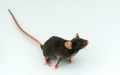
Humanized immune system (HIS) mice have a variety of applications for studying diseases in which the immune system drives pathogenesis. This includes immuno-oncology, autoimmunity, and a less discussed but also crucial subset of immunologic disorders, allergy. Indeed, Taconic's CD34+ hematopoietic stem cell (HSC)-engrafted NOG-EXL (
huNOG-EXL) is a model that supports the key myeloid cell populations that mediate allergic responses in humans.
Humanized NOG-EXL mice support key myeloid cell populations
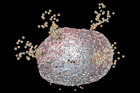
The humanized NOG-EXL mouse, engrafted with CD34+ HSC, was originally characterized in allergic disease. The NOG-EXL mouse transgenically expresses human GM-CSF and human IL-3. These cytokines support the development and maintenance of human myeloid cells after engraftment, including granulocytes, monocytes, mast cells, basophils, neutrophils, and dendritic cells. Like the humanized NOG mouse upon which the huNOG-EXL is based, these mice also support T and B cells. The presence of myeloid cells — especially mast cells, which will be discussed in the case studies outlined below — is what makes the huNOG-EXL a useful model for studying allergic disease.
Human myeloid cells respond to GM-CSF and IL-3 signaling
Mast cells, basophils, eosinophils, and neutrophils are key cell populations driving allergic reactions in humans and humanized immune system mice. All of these populations can be found in the huNOG-EXL, where CD33+ cells comprise 5-20% of the hCD45+ cell population. Human mast cells, eosinophils, and basophils have all been reported to express receptors for both hGM-CSF and hIL-3, which are transgenically expressed by the NOG-EXL
1. Neutrophils express the receptor for hGM-CSF, and engagement of this receptor upregulates expression of the IL-3 receptor
2.
The impact of IL-3 and GM-CSF on mast cell function in vivo
Interestingly, while murine mast cells rely on IL-3 for early differentiation from progenitor cells, this is debatable with regard to human mast cells
3,4. In humans, IL-3 and GM-CSF typically promote differentiation into granulocytes. Human mast cell progenitors that have seen IL-3 or GM-CSF
in vitro were shown in one study to retain the ability to differentiate into mast cells but only express low levels of the mast cell marker c-Kit
3. Instead, IL-3 appeared to specifically support mast cell precursor cell replication and survival
3,5. Conversely, another study indicated that IL-3 is sufficient for differentiation of myeloid precursors to the pre-mast cell stage expressing c-Kit (Dahlin 2017). One may hypothesize that
in vivo, such as in HIS mice, other immune cell populations could be responding to hIL-3/hGM-CSF by producing cytokines such as IL-6 to drive terminal mast cell differentiation
6-8.
Case Studies
Mast cells mediate Type I hypersensitivity and anaphylaxis
The original publication characterizing the CD34+ HSC-engrafted NOG-EXL described its utility in Type I hypersensitivity reactions using the pollen challenge to model anaphylaxis. The group from the Central Institute for Experimental Animals (CIEA) concluded that the NOG-EXL engrafted with a human immune system supported functional tissue-resident mast cells that can mediate allergic responses following sensitization. These mast cells were characterized as FcεRI+ cells expressing mast cell chymase and tryptase. In humans, these cells are localized to the skin; in the humanized NOG-EXL, they are found in the skin as well as the spleen, lung, and stomach
9. NOG-EXL engrafted with CD34+ HSC can therefore support tissue-resident human mast cells that can mediate cutaneous allergic reactions.
Cytokines produced by ILC2 and mast cells drive asthma
A follow-up study by the group that initially characterized the humanized NOG-EXL then analyzed its utility as a model for asthma. While there are many murine asthma models available, most do not have all the key characteristics of human disease such as the infiltration and impact of human myeloid cells in the airway. Thus, the huNOG-EXL, with its support for eosinophils, mast cells, and basophils in addition to leukocytes, recapitulates this feature of human asthma in a unique way. Infiltration of human T cells, eosinophils, mast cells, and basophils is observed in the bronchial alveolar lavage fluid (BALF) of IL-33-treated humanized NOG-EXL. This inflammation can be ameliorated by blocking IL-13, a cytokine produced by ILC2, T cells, and mast cells that drives airway inflammation downstream of IL-33
10. Humanized NOG-EXL animals therefore encompass more features of human asthma than previous HIS models used to study this disease.
Humanized NOG-EXL mice can be sensitized to food allergens
The same group most recently used CD34+ humanized NOG-EXL mice to develop a model of anaphylactic food allergy. Upon challenge with an oral antigen to which they had previously been sensitized, expression of the human cytokines MCP-1, IFNg, IL-6, and IL-8 increase, however, the levels of mouse cytokines were not impacted. This demonstrates the utility of this model for studying human food allergy without interference from the residual mouse immune system. Humanized NOG-EXL also exhibit a rapid decrease in body temperature and succumb to anaphylaxis within 1 hour following challenge, whereas humanized NOG mice (which do not express hGM-CSF or hIL-3) do not. In parallel, humanized NOG-EXL mice have increased serum histamine and increased expression of the mast cell degranulation marker CD63, both hallmarks of an anaphylactic response mediated by mast cells
11.
Future directions for using huNOG-EXL to study allergy
The Taconic team is eager to expand the use of huNOG-EXL to study allergic diseases. While the proof-of-concept studies have been published for close to a decade, relatively few publications have taken advantage of the expanded myeloid compartment in the
huNOG-EXL to study allergy. The presence of functional human mast cells as well as basophils, eosinophils, and neutrophils make this an enticing model for these acute immune responses.
 Download the Taconic Biosciences White Paper:
If you are interested in discussing a potential collaboration with Taconic to generate data in this area, please reach out to schedule a consult with our Field Application Scientist team.
References:
1. Dahl, C.; Hoffmann, H. J.; Saito, H.; Schiøtz, P. O. Human Mast Cells Express Receptors for IL-3, IL-5 and GM-CSF; a Partial Map of Receptors on Human Mast Cells Cultured in Vitro. Allergy 2004, 59 (10), 1087-1096.
2. Smith, W. B.; Guida, L.; Sun, Q.; Korpelainen, E. I.; Heuvel, C. van den; Gillis, D.; Hawrylowicz, C. M.; Vadas, M. A.; Lopez, A. F. Neutrophils Activated by Granulocyte-Macrophage Colony-Stimulating Factor Express Receptors for Interleukin-3 Which Mediate Class II Expression. Blood 1995, 86 (10), 3938-3944.
3. Hjertson, M; Sundström, C; Nilsson, K; Nilsson, G. The Potential of Human Mast Cell Progenitors to Differentiate into Mature Mast Cells Remains after Prolonged Culture with Flt3 Ligand, Interleukin-3 or Granulocyte-macrophage Colony Stimulating Factor. Brit J Haematol 1999, 104 (3), 516-522.
4. Dahlin, J. S.; Ekoff, M.; Grootens, J.; Löf, L.; Amini, R.-M.; Hagberg, H.; Ungerstedt, J. S.; Olsson-Strömberg, U.; Nilsson, G. KIT Signaling Is Dispensable for Human Mast Cell Progenitor Development. Blood 2017, 130 (16), 1785-1794.
5. Brandt, J. E.; Bhalla, K.; Hoffman, R. Effects of Interleukin-3 and c-Kit Ligand on the Survival of Various Classes of Human Hematopoietic Progenitor Cells. Blood 1994, 83 (6), 1507-1514.
6. Sonderegger, I.; Iezzi, G.; Maier, R.; Schmitz, N.; Kurrer, M.; Kopf, M. GM-CSF Mediates Autoimmunity by Enhancing IL-6-Dependent Th17 Cell Development and Survival. J Exp Medicine 2008, 205 (10), 2281-2294.
7. Desai, A.; Jung, M.-Y.; Olivera, A.; Gilfillan, A. M.; Prussin, C.; Kirshenbaum, A. S.; Beaven, M. A.; Metcalfe, D. D. IL-6 Promotes an Increase in Human Mast Cell Numbers and Reactivity through Suppression of Suppressor of Cytokine Signaling 3. J Allergy Clin Immun 2016, 137 (6), 1863-1871.e6.
8. McHale, C. C.; Deppen, J.; Shirley, D.; Gomez, G. Human Skin Mast Cells Constitutively Express Gp130 and Functional IL-6 Receptor | The Journal of Immunology. J Immunol 2016, 196 (1 Supplement).
9. Ito, R.; Takahashi, T.; Katano, I.; Kawai, K.; Kamisako, T.; Ogura, T.; Ida-Tanaka, M.; Suemizu, H.; Nunomura, S.; Ra, C.; Mori, A.; Aiso, S.; Ito, M. Establishment of a Human Allergy Model Using Human IL-3/GM-CSF-Transgenic NOG Mice. J Immunol 2013, 191 (6), 2890-2899.
10. Ito, R.; Maruoka, S.; Soda, K.; Katano, I.; Kawai, K.; Yagoto, M.; Hanazawa, A.; Takahashi, T.; Ogura, T.; Goto, M.; Takahashi, R.; Toyoshima, S.; Okayama, Y.; Izuhara, K.; Gon, Y.; Hashimoto, S.; Ito, M.; Nunomura, S. A Humanized Mouse Model to Study Asthmatic Airway Inflammation via the Human IL-33/IL-13 Axis. Jci Insight 2018, 3 (21), e121580.
11. Ito, R.; Katano, I.; Otsuka, I.; Takahashi, T.; Suemizu, H.; Ito, M.; Simons, P. J. Bovine β-Lactoglobulin-Induced Passive Systemic Anaphylaxis Model Using Humanized NOG HIL-3/HGM-CSF Transgenic Mice. Int Immunol 2020.
 Humanized immune system (HIS) mice have a variety of applications for studying diseases in which the immune system drives pathogenesis. This includes immuno-oncology, autoimmunity, and a less discussed but also crucial subset of immunologic disorders, allergy. Indeed, Taconic's CD34+ hematopoietic stem cell (HSC)-engrafted NOG-EXL (huNOG-EXL) is a model that supports the key myeloid cell populations that mediate allergic responses in humans.
 Humanized immune system (HIS) mice have a variety of applications for studying diseases in which the immune system drives pathogenesis. This includes immuno-oncology, autoimmunity, and a less discussed but also crucial subset of immunologic disorders, allergy. Indeed, Taconic's CD34+ hematopoietic stem cell (HSC)-engrafted NOG-EXL (huNOG-EXL) is a model that supports the key myeloid cell populations that mediate allergic responses in humans.






.jpg)

.jpg)
.jpg)
.jpg)
.jpg)


.jpg)


.jpg)
.jpg)

.jpg)


.jpg)


.jpg)

.jpg)


