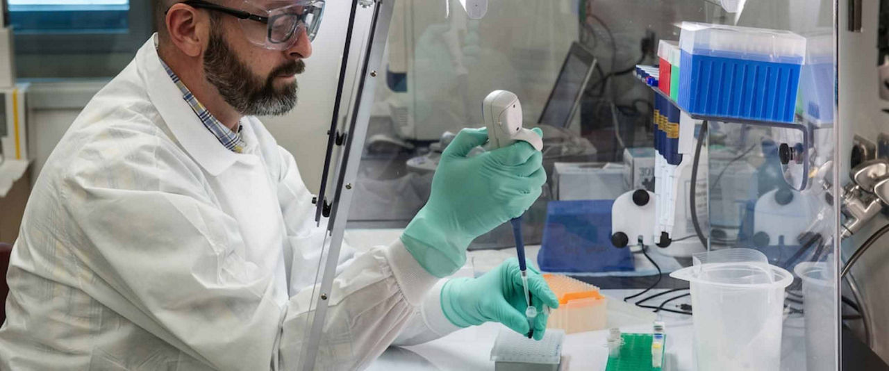
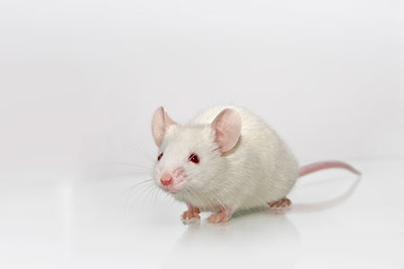
 Dr. Janell Richardson recently presented a webinar on a case study that explored variance and its implications on study design in the huNOG-EXL, a humanized immune system (HIS) model. HIS models are the only in vivo model types to date that allow investigators to study, in real time, human immune cell function under both physiological and pathological conditions.
Dr. Richardson provided an overview of HIS mice and the factors that can impart variance when using HIS models like the huNOG-EXL. She then presented a case study based on a Taconic Biosciences R&D study using huNOG-EXL in an immuno-oncology application, reviewing factors that impact reconstitution, immuno-oncology utility of the model, and study design.
Following her presentation, Dr. Richardson addressed many questions from webinar participants. Here we present the full webinar Q&A.
Q: Can you comment on humanization with pooled cells from different donors?
Dr. Janell Richardson: In the early days of Taconic humanizing mice, we asked that question ourselves: pooled donors vs. single donors. A generality of the commercial space is that we tend to move away from pooled donors, because the number of variables that end up being contained within a mouse really heavily influences those engraftment kinetics that we're talking about. The degree of failure can go up. Remember how I highlighted that standard deviation of 20 and a failure of less than 10? We would definitely expect both of those to go up if we were using pooled donors. For us to mitigate that risk, we use single donors. And I know that many others that engraft their own animals follow the same type of general thought process. But I can't say, "show X and Y" and then tell you why or why not. It's your own experiences and what works for you. What we find works best for us is to use single donor sources for the reasons I highlighted.
Q: Do you have a recommended timeline to inject patient-derived xenograft (PDX) tumors after CD34+ HSC engraftment so you can avoid those macrophage issues but still get enough human cells?
JR: We planned for what we needed to, but I would say we did not execute the full plan the way that I would have ideally liked. Unfortunately, the vast majority of this data happened just as COVID was hitting the world. For our own models off the shelf, we QC these at 10 weeks post-engraftment, so that means typically they come to your door between 12-13 weeks post-engraftment. I'd say get them in and acclimated, make sure they're comfortable, and then I would make sure you plan your PDXs. Which means that your warm tumors have to be ready to go probably by about two weeks after your delivery to your door. Our warm tumors were ready too far in advance for when our animals were coming in. You have to align that with your animal delivery and the necessary acclimation. So once your PDXs come on board, you should know your profile in terms of your time and kinetics, and as soon as that meets that criteria, I would go ahead and randomize them to the group and apply your therapeutics. I really would argue that the tighter you can move up to that timeline, it gives you more room for error. Because there really is a piece here where the donors can influence the study window itself. We have some donor-engrafted huNOG-EXLs that can last for 48 weeks post-engraftment. We have others that start to go downhill at 22 weeks post-engraftment. There's no way to predict.
Q: What would be a minimum number of donors and what would be an ideal number of donors?
JR: There's no definitive answer. I can't point you to a publication that says you should do this or that. It's all based on the type of question you're asking, the degree of risk you're willing to take, and the clarity and granularity that you need to answer that question. Two black/white scenarios are: I just need to know if this tumor grows in this model, vs. a first-in-human dosing paradigm. I would definitely not do an N of 1 for a first-in-human potential dosing paradigm. Would I do an N of 1 for tumor growth? Possibly. It depends on the tumor. If it's glioblastoma, and maybe I'm putting it subcutaneous and I know it grows very quickly, I might just do an N of 1 donor. In terms of a dosing paradigm, the minimum that I feel comfortable with is an N of 3 donors. Depending on the risk, I would say that kind of controls the number of donors. I would start to look at maybe 5 donors if I'm really asking that critical question: whether I'm going to carry this drug through, whether I'm going to take the next steps.
Q: You showed us data on human cells in peripheral blood and spleen. Do you have information on the distribution of human immune cells in different tissues, such as the gut, in huNOG-EXL mice?
JR: To my knowledge we do not yet have that data, but we very much want that data. Those are some goals for us over the next year. We partner with different entities and we get some very good pieces of information, but we have to tackle and take off bits and pieces. Understanding tissues such as the lung, the skin, and the gastrointestinal tract in these animals is definitely of interest to us. We have some more recent data that gives us maximum granularity on the peripheral blood and spleen. We're hoping that in the next couple of months we're able to publicly release that as well. It was using CyTOF Our goal is to increase the level of data that we have so that you consult with us. I would say, really reach out, and keep reaching out, because that changes on a month-to-month basis.
Q: Is it possible to induce colitis in huNOG-EXL mice, and if so, what type of colitis model?
JR: The answer is, stay tuned. We have an active collaboration going on right now. We looked at one last year, I believe that was a model of DSS induced-based colitis in the huNOG-EXL, and we were unsuccessful with that endeavor. We have another type of colitis model that we're looking to ask the question in the huNOG-EXL with a partner.
Q: Do you have any data on orthotopic PDX implantation in humanized mice? And do you know if TIL infiltration is variable depending on the orthotopic organ of interest?
JR: I believe there was a webinar given in December by an organization that really would have a very good understanding of this, called Certis. To my knowledge, they are looking at the question in terms of subcutaneous vs. orthotopic in un-engrafted models and then will move to engrafted models. I can say that glioblastoma is probably the PDX that I am most familiar with in terms of orthotopic vs. subcutaneous, and I can say that orthotopically does show infiltration that does differ vs. the subcutaneous application of a glioblastoma, which grows quite well in the NOG-EXL. Unfortunately, that data is still confidential, but if we're able to get it publicly released we will do so. Unfortunately, the answer is, not at this time, but stay tuned. We're hoping to have a public asset to that by the end of this year.
Q: Can you talk about some of the general characteristics in human immune cell reconstitution and kinetics in the huNOG-EXL? And how does that compare to another model that can be used for myeloid cell studies: the human IL-6 NOG model?
JR: Hopefully within a couple of months we'll really be able to give you the "why" to my answer. Because the CyTOF that was done in partnership with a big pharma entity was actually using the same donor-engrafted NOG-EXL compared to hIL-6 NOGs. So, it is able to be directly compared, although the time profile is a little bit different. There are two important pieces that we have taken away in terms of the NOG-EXL vs. the hIL-6 NOG. I would say the differences are minimal compared to the similarities. In terms of some of the similarities: They both show myeloid reconstitution when you engraft hematopoietic stem cells into them. The NOG-EXL engrafts still at a much higher overall percentage and efficiency compared to the IL-6 though. For example, if I were to engraft animals and take a snapshot at 12 weeks post-engraftment, my expectation would be CD45+ in the NOG-EXL somewhere around 65%. I would expect CD45+ in the hIL-6 NOG to be somewhere around 45%. It doesn't mean that it stays there, but again, it's a general idea of similarities vs. differences. Another piece is IL-6 certainly is known for its influence on B cells. We have seen, although it can be very donor dependent, the potential for a very large B cell reconstitution profile compared to the NOG-EXL. But again, it is heavily dependent on the donor that it does not carry through all the way. Another difference would be Tregs (regulatory T cells). NOG-EXLs typically have a higher percent of Tregs than the hIL-6 NOGs do. As opposed to the actual myeloid reconstitution, typically between 8-12% is seen in both the peripheral blood and the spleen of both the NOG-EXL and the hIL-6 NOG. So, they are very compatible in terms of myeloid differences, granulocytes. We don't know for sure the degree of granulocytes. In the hIL-6 NOG we know that they are there, but they're not to the level that they are in the NOG-EXL. The hIL-6 NOG manuscript that was published by our partner, CIEA, used it in a head and neck cancer-based model and showed its increase in TAMs (tumor-associated macrophages) and MDSCs (myeloid-deprived suppressor cells). We have not been able to use it yet in a cancer model, so I can't speak to potentially its immuno-oncology based utility differences.
Q: I heard you say the huNOG-EXL would not be a model you would recommend for NK cell questions. Can you talk about what some of the options might be?
JR: I would say it could be appropriate for NK cells if it's additional. If it's your primary cell of interest, then it's not the NOG-EXL. What would be a model I would choose? In the chart, there are two models you could choose: either the hIL-2 NOG or the hIL-15 NOG. The hIL-2 NOG study window is much, much shorter typically than the hIL-15 NOG study window. Most individuals who are concentrating on NK cells typically follow a creation protocol utilizing a human hIL-15 NOG that is irradiated and engrafted with purified and isolated NK cells from human peripheral blood. So, you typically can see persistence. I think the ideal is somewhere around three weeks post-engraftment and you get these nice, clean NK cells as well as the persistence, which is a big one with NK cells. You can get them into the NOG-EXL, but often in models they disappear, like, snap!
Q: How does the huNOG-EXL compare to the huNSG-SGM3 model, which is another model commonly used for myeloid ell applications?
JR: A lot is said in terms of NOG, NSG, NRG. The field typically lumps them all into a super-immunodeficient strain, and for the most part, they're fairly biologically equivalent, especially the NOG and the NSG. The differences, for what we sometimes refer to as the double humanized--in that they are genetically humanized plus they have the human cells that are transplanted into them--that's where the strain and the vendor very much matter. The vendor can still matter for super-immunodeficients as well, and that gets to microbiome and environmental issues, but going to the utility and application, human transgene cytokine expressing models very much differ from each other. There are no biological equivalents. Each one is very different from each other. Pieces to look at to separate them: What is the cytokine expression level? We make an IL-15, Jax makes an IL-15. They differ in terms of their plasma IL-15 concentration. I'm going to reciprocate that for the question you asked specifically: the NSG-SGM3 and the NOG-EXL. They not only have very different cytokine expression levels, but they actually express different genes. The NOG-EXL shares two genes (it only has two): IL-3 and GM-CSF, and the NSG-SGM3 has three (SCF, GM-CSF, and IL-3). Most importantly, those cytokines are in very, very different levels. We have an Insight on this on our website. I strongly encourage you to look at it. There's actually a PDF that goes into the comparisons to different myeloid-expressing super-immunodeficient models such as the MISTRG and now there's the MISTRG6, which of course has IL-6 added to it. But really the important part is, what is the advantage and what is the disadvantage? I'm getting something, but you're always taking something away. It's never as easy as, this model has everything I need it to do and is spectacular and will last forever. If someone says that, then that's a new model to my understanding. Our NOG-EXL has a much, much lower cytokine expression level than the NSG-SGM3. Why is that important? It's important because of the study window. The study window is dramatically different between the two models. Also, the reconstitution and the kinetics are drastically different between the two models. If you want a model, and you only need it to be around for two or three weeks, and you're looking at T cells, the NSG-SGM3 is a rocket ship for T cells, especially Tregs, but it comes at a price. The price is HSC exhaustion, and the model can't live for very long with a huge amount of human T cells, as well as macrophages: macrophage-activation syndrome, hemophagocytic lymphohistiocytosis (HLH), the animal succumbs. It doesn't mean that the NOG-EXL is immune to that. All myeloid expressing models can suffer from macrophage-activation syndrome. The degree to which they suffer from it, and the time to onset, differs for every single one of those. That is a key difference.
Schedule Consultation







.jpg)

.jpg)
.jpg)
.jpg)
.jpg)



.jpg)


.jpg)
.jpg)


.jpg)


.jpg)

.jpg)


