What is a Xenograft?

by Caroline Horizny Mitchell, PhD | Updated: December 23rd, 2024

Key TakeawaysKey Takeaways

  • The term xenograft refers to tissue or an organ originating from a species different from that of the recipient. Xenografts are invaluable tools for oncology research and play a crucial role in clinical wound treatment.
  • Numerous mouse and rat PDX models have been developed to accept patient-derived xenografts. To prevent the rejection of the foreign tissue, these models are typically severely immunodeficient or contain a human immune system.
  • The advent of humanized mice has introduced even more options to the mouse model landscape — allowing researchers to engraft tumors to a mouse that contains a human immune system.


The term xenograft refers to a tissue or organ that is derived from a species that is different from the recipient of the specimen. They are powerful research tools in oncology as well as essential for treating wounds in the clinic.

This Insight will highlight a few key uses of xenografts and why they are important to research studies.

Xenografts in the Treatment of Skin Injuries

In the event that a person is very badly burned or injured and is lacking large areas of skin, xenografts are used to temporarily repair the affected areas. The most commonly used xenograft is the EZ Derm®, which is an aldehyde cross-linked porcine dermis that aids in the recovery of partial-thickness skin loss. It is not a permanent treatment but provides coverage to the affected area long enough to protect the exposed tissue from external contaminants and decrease protein loss and cell death.

At the time of injury, autografts are not always feasible treatment options depending on the size and location of the skin injury. Once the skin underneath the xenograft has healed to an acceptable state, a thin layer of skin may be taken from an unaffected location and used to cover the tissue injury. However, if not enough donor skin is available a meshed graft will need to be used, wherein the donor skin is stretched and sliced to create a larger mesh-like covering1. Recovery from this graft is more difficult and takes longer, but both options have proved successful in the clinic2.

Patient-Derived Xenografts (PDX) for Oncology Research

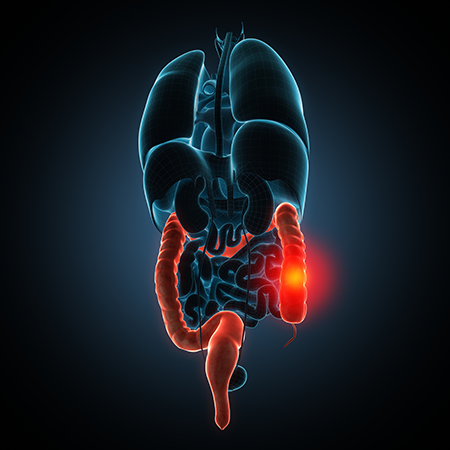
A second use for xenografts is in oncology research. In order to develop a personalized treatment plan for cancer patients, a small segment of their tumor may be excised and subsequently grafted into an immunodeficient or humanized mouse. These are referred to as patient-derived xenografts (PDX)3.

In addition to personalized treatments, PDX models allow for the study of the tumor and its natural growth patterns and behavior. Depending on the tumor's original location it can be transplanted under the skin or into the organ that the tumor was originally derived from.

Immunodeficient Mouse Models for PDX studies

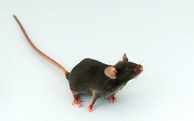
Numerous mouse and rat PDX models have been developed to accept patient-derived xenografts. To prevent the rejection of the foreign tissue, these models are typically severely immunodeficient or contain a human immune system.

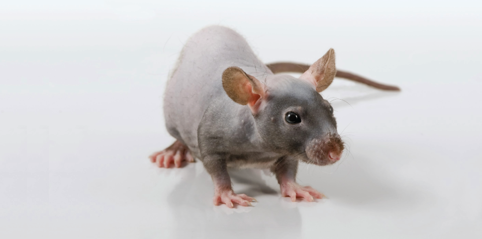
Both nude mice (which lack T cells) and scid mice (which lack T and B cells) have been used for PDX studies4. This nature of immunodeficiency allows for the xenograft to be accepted and not attacked by host immune responses5. Genetically engineered mice (GEMs) also present viable options for xenograft studies because of their modified genomes resulting in a lack of B and T cells. One specific example is the mouse model with a disruption in the Rag2 gene. Mice that are homozygous for this modification are unable to initiate V(D)J recombination resulting in the failure to produce mature B and T lymphocytes6.

While all of these models are considered immunodeficient, the field of PDX studies has recently moved towards using even more immunodeficient models such as the CIEA NOG mouse®. This model in particular provides a better platform for the engraftment and growth of patient-derived tumors.

This GEM was originally developed by Mamoru Ito of the Central Institute for Experimental Animals (CIEA) in Japan. This model lacks mature T, B, and NK cells, displays reduced complement activity, has dysfunctional macrophages and dendritic cells, and does not display 'leakiness' of T or B cells over time. With all of these characteristics, this model has been shown to be an excellent platform for xenograft studies7.

Humanized Mice

The advent of humanized mice has introduced even more options to the mouse model landscape — allowing researchers to engraft tumors to a mouse that contains a human immune system. Multiple models with these modifications are based on the NOG background and have been engrafted with human hematopoietic stem cells.

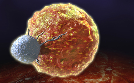
After stable engraftment of human hematopoietic stem cells, human lymphocytes will be present in peripheral blood, bone marrow, the thymus, and the spleen. These models present unique and invaluable platforms for studying novel immunotherapies on patient-derived tumors.

References:

  1. Skin grafting. Accessed May 13, 2019.
  2. Fatah, M. F.; Ward, C. M. The Morbidity of Split-Skin Graft Donor Sites in the Elderly: The Case for Mesh-Grafting the Donor Site. Br. J. Plast. Surg. 1984, 37 (2), 184-190.
  3. Lai, Y.; Wei, X.; Lin, S.; Qin, L.; Cheng, L.; Li, P. Current Status and Perspectives of Patient-Derived Xenograft Models in Cancer Research. J. Hematol. Oncol. 2017, 10 (1), 1-14.
  4. Blunt, T.; Finnie, N. J.; Taccioli, G. E.; Smith, G. C.; Demengeot, J.; Gottlieb, T. M.; Mizuta, R.; Varghese, A. J.; Alt, F. W.; Jeggo, P. A.; et al. Defective DNA-Dependent Protein Kinase Activity Is Linked to V(D)J Recombination and DNA Repair Defects Associated with the Murine Scid Mutation. Cell 1995, 80 (5), 813-823.
  5. Richmond, A.; Su, Y. Mouse Xenograft Models vs GEM Models for Human Cancer Therapeutics. Dis. Model. Mech. 2008, 1 (2-3), 78-82.
  6. Shinkai, Y.; Rathbun, G.; Lam, K. P.; Oltz, E. M.; Stewart, V.; Mendelsohn, M.; Charron, J.; Datta, M.; Young, F.; Stall, A. M. RAG-2-Deficient Mice Lack Mature Lymphocytes Owing to Inability to Initiate V(D)J Rearrangement. Cell 1992, 68 (5), 855-867.
  7. Ito, M.; Hiramatsu, H.; Kobayashi, K.; Suzue, K.; Kawahata, M.; Hioki, K.; Ueyama, Y.; Koyanagi, Y.; Sugamura, K.; Tsuji, K.; et al. NOD/SCID/ Ɣcnull Mouse: An Excellent Recipient Mouse Model for Engraftment of Human Cells. Blood 2002, 100 (9), 3175-3182.

Related Resources

On-Demand Webinar

Patient-Derived Xenografts on Humanized Mice

In this exclusive Taconic Biosciences webinar, learn how to develop PDX models of cancer on humanized mouse models, unlocking the ability to study real human cancers in the context of a functional, humanized immune system.

Webinar

On-Demand Webinar

Estrogen-Related Effects in Breast Cancer Xenograft Models

In this webinar, we will explore the impacts of external estrogen supplements on various strains of mice used as breast cancer xenograft models in preclinical research.

Webinar

On-Demand Webinar

Next Generation Humanized Mice for Acute Myeloid Leukemia and Natural Killer Cell Therapy Testing

In this webinar, Amy Wesa, PhD, of Champions Oncology Inc, will discuss how complementing these translational PDX models with the introduction of human immune cells is driving the next generation of patient-derived models.


Get In Touch

Book a complimentary consultation

Taconic Biosciences' model generation team has produced about 5,000 models in the last 15 years, developing a globally-recognized reputation for advancing the work of in vivo researchers. Our scientific program managers are here to help you navigate the complexities of model generation.

If you need immediate assistance, please contact Customer Service:

Taconic Corporate Offices

Email: info@taconic.com

Phone: +1 (518) 697-3900

273 Hover Ave., Germantown, NY 12526

North American Customer Service

Email: info@taconic.com

Phone:
+1 (518) 697-3915 

Toll-free:
+1 (888) 822-6642


Hours:
(Monday - Friday): 7 a.m. - 6 p.m. ET

European Customer Service

Email: info@taconic.com

Phone (Europe and Denmark):
+45 70 23 04 05 

Phone (Germany):
+49 214 50 68 023 

Hours: (Monday - Friday):
7 a.m. - 5 p.m. CET