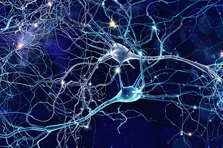
 Parkinson's Disease (PD) affects over 10 million people worldwide and is the second most common neurodegenerative disorder1,2. Key research areas include genetics studies of patients with PD, target optimization for new treatments, the biology of cellular signaling pathways (α-synuclein, LRRK2, GBA, PRKN/PINK1, inflammation, and mitochondrial dysfunction), biomarkers for PD onset and progression, and new imaging techniques to measure brain changes and activity.
The Parkinson's Progression Markers Initiative (PPMI)
The Parkinson's Progression Markers Initiative (PPMI) was launched in 2010 with the goal of identifying new biomarkers involved in PD development and progression. As of 2018, the PPMI completed the enrollment of 1,400 participants with 600 rare genetic mutations. A critical part of this initiative is in the collection, storage, and distribution of PD biosamples. The study has established the most robust database of PD specimens and associated data and has received over 100 specimen requests thus far3.Role of Inflammation in PD
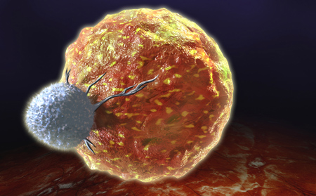
Inflammation in the brain, known as neuroinflammation, plays a crucial role in the development and progression of PD. In the brain of PD patients, over-activated microglia and astrocytes have been shown to release a number of pro-inflammatory cytokines, thereby promoting neuroinflammation4. Interestingly, multiple studies have been published on the effects of chronic nonsteroidal anti-inflammatory (NSAID) drug regimes in decreasing the incidence and progression of neuroinflammation in PD5.Biomarkers for Inflammation in PD
Biomarkers are biological markers that can be used to accurately and reproducibly diagnose disease onset or monitor response to treatment. Biomarkers are directly related to molecular pathways, and the more closely related to a specific condition, the better clinicians can predict and diagnose disease onset and progression.In PD, the use of inflammation as a biomarker can be studied to inform treatment regimes. Inflammation can be used in two ways: to determine if a person might be prone to developing PD, and to assess the efficacy of treatment of symptoms of PD.
Pathways in PD
Inflammation is a complex process with multiple molecular pathways contributing to its onset and progression. There are several ways to monitor neuroinflammation with the most common technique being brain scans through PET imaging. Specific markers identified through a PET scan can detect neuroinflammation in the central nervous system (CNS). However, markers of neuroinflammation may also present themselves in the periphery (blood, urine, saliva, etc.), which would enable the study of PD progression in a less-invasive manner.Molecular Assays to Detect Inflammation Biomarkers in PD
The PPMI has funded a number of projects to identify inflammation biomarkers that could potentially be used in a non-invasive diagnostic assay for PD, several of which have been supplemented for follow-on funding. While these projects required the use of human samples and enrolled both healthy and PD patients in clinical studies, rodent models have been essential to the progression of research in this field. These animal models are often directed at elucidating the involvement of specific disease pathways.Mouse Models for PD Studies
Two key mouse models used for studying PD are the Rab29 Overexpression Mouse and the LRRK2 Targeted Replacement Mouse. The LRRK2 mouse includes a human G2019S point mutation in exon 41 of the mouse LRRK2 gene. The G2019S alpha-mutation has been linked to the development of both sporadic and familial PD6. In patients, the G2019S LRRK2 mutation is an autosomal dominant mutation that leads to a pathology that is similar to that observed in idiopathic PD. The Rab29 mouse model adds an additional benefit to PD researchers due to the involvement of the Rab29 GTPase in stimulating LRRK2 activity7. Together, both play a role in vesicle trafficking and maintaining the endosome-trans-Golgi network structure.| Model Name | Model Number | Mutation |
| Rab29 Overexpression Mouse | Model 16552 | Constitutive knock-in of murine Rab29 under control of the Pgk promoter generated by targeted transgenesis into the ROSA26 locus. |
| Human LRRK2 G2019S Targeted Replacement Mouse | Model 13940 | Contains the human G2019S point mutation introduced into exon 41 of the mouse LRRK2 gene. Additionally, exon 41 is flanked by lox P sites, potentially allowing generation of a LRRK2 gene constitutive KO model upon intercrossing with a Cre deleter strain. |
Role of α-synuclein in PD
Many GEMs have been created to overexpress the human α-synuclein (αSyn) protein, including key models such as the Human Alpha Synuclein Rat, Human Alpha Synuclein A53T Rat, and Human Alpha Synuclein E46K Rat. While multiple research groups have successfully used these models for PD research, consistency in experimental results remains a key issue. Utilizing αSyn pre-formed fibrils (αSyn PFFs) is the main focus of study right now, as these types of models extend pathology and applications beyond previous transgenic αSyn models8. To better replicate and understand the implications of experimental results, the microbiome must be considered, as the microbiome is increasingly cited as a potential contributor to neurological conditions and neurodegenerative diseases.Microbiome Influence on PD Development and Progression
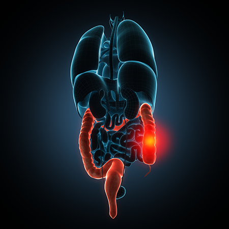
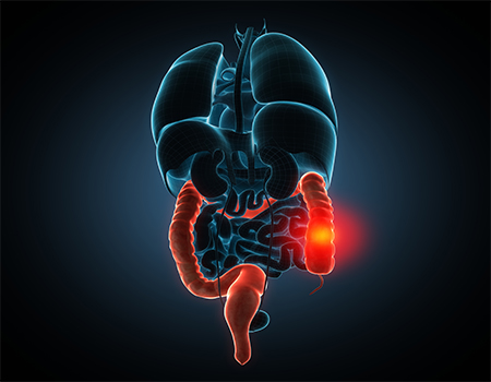
In 2006, Heiko Braak proposed a physical connection between the gut and the brain resulting in αSyn transport between locations9. Since then, multiple studies have been published linking the development and progression of PD to the gut-brain axis, as well as the transfer of αSyn through the vagus nerve to the brain10-12.The composition of the gut microbiome can serve as both an early biomarker and a potential therapeutic option when treating PD. The microbiome and inflammation pathways are intimately connected, as evidenced by an unbalanced gut microbiota and the development of inflammatory conditions such as inflammatory bowel disease (IBD).
In a paper published in Neuron in 2019, a research team led by Ted Dawson from the Johns Hopkins School of Medicine, tested Braak's initial hypothesis in a novel gut-brain αSyn transmission mouse model. In brief, the researchers injected mouse αSyn PFF into the muscle layers of the pylorus and duodenum, as these locations are highly innervated by the vagus nerve13. Mice were monitored for up to 10 months after this injection and immunoblot analysis over the course of the study indicated that αSyn was found in the medulla oblongata, pons, amygdala, ventral midbrain, hippocampus, stomach, and duodenum. A significant loss of dopaminergic neurons was also observed. Multiple tests were conducted to assess the behavioral response to spatial learning, fine motor dexterity, cognition, and emotional state. When compared to controls, the experimental mice displayed multiple cognitive impairments such as memory loss, social deficits, anxiety, depression, and disruptions in normal gastrointestinal function14.
Taken together, these research efforts demonstrate a crucial role for the microbiome on the development and progression of PD. Mouse and rat models are expected to be critical tools in continued research on the role of microbiome in PD as well as other aspects of this disease.






.jpg)

.jpg)
.jpg)
.jpg)
.jpg)



.jpg)


.jpg)
.jpg)

.jpg)


.jpg)


.jpg)

.jpg)


