CRE-Luc GPCR Reporter Mouse Platform
Features:
- A panel of luciferase reporter mice to assay GPCR ligand binding and pathway activation in various tissues.
- Tissue specific expression (eg., CNS, pancreas etc.) of CRE-Luc reporter.
- Same reporter system supports in vitro, in vivo, or ex vivo assays
- Ease the in vitro to in vivo transition and provide better selection of optimized active leads.
- The platform can provide rapid in vivo PK/PD profiling of compounds with quantitative data to compare pharmacological action
- Assay can be used for two main GPCR classes, Gs and Gi
- The CRE-Luc transgenic models contain a luciferase reporter under the control of a synthetic promoter CRE, which supports real-time bioimaging of GPCR ligand activity.
- GPCR signaling through the cAMP pathway can be detected from the target GPCR that is in a native cellular environment with a full complement of associated receptors and membrane constituents.
- Forskolin stimulation will increase cellular cAMP levels and increase the luciferase signal derived from the CRE-Luc transgene
- Specific GPCR agonist will:
- Increase the luciferase signal for Gs or Gq coupled receptors
- Decrease the luciferase signal for Gi coupled receptors
- Specific GPCR antagonist or inverse agonist will:
- Decrease the luciferase signal for Gs or Gq coupled receptors
- Increase the luciferase signal for Gi coupled receptors
- The Cre-Luc platform accelerates the transition from HTS to in vivo profiling of GPCR small molecule leads as well as defines the mode of action of GPCR drugs.
- The Cre-Luc platform supports in vitro, in vivo, and ex vivo assays helping with the in vitro to in vivo transition of assays.

The CRE-Luc lines can serve as a source of primary cells with the GPCR reporter in its native environment. Therefore in vitro studies can be first performed followed by in vivo studies.
In vitro Studies (Primary Cells and ex vivo assays)
Initial analysis of ligand activation can first be confirmed in CRE-Luc primary cell cultures. For example, CRE-Luc cell cultures support GPCR receptor specificity assays, e.g., through the use of RNAi or ligand competition assays; these are an important validation step since potentially any receptor (or combination of receptors) can be activated by a single ligand.
After profiling ligand activation in primary cells, more complex tissue profiles can assayed either ex vivo or through isolated tissue homogenates that assay luciferase enzyme levels. Tissue homogenate analysis is time consuming, but when used with compound dosing in whole animals, can generate a detailed, tissue specific, and quantitative ligand activation profile.

Images of compound induced changes in luciferase level by a β-adrenergic receptor agonist.
In vivo Studies (Whole Animal)
After activation profiles are established from primary cells and tissues, then ligand profiles can be assayed in whole animals by bioimaging and incorporating dose response and time course assays. Data analysis can occur in the same day as the imaging session which allows unknown endpoints or results in the assay to be defined as the study progresses. This feature impacts flexibility in the animal study and can save significant time in avoiding repetitive studies to capture overlooked data. The whole animal bioimaging assay format can quantitatively define the site and magnitude of ligand activation plus support a quantitative comparison of similar compounds for selection of optimal lead structures and SAR.

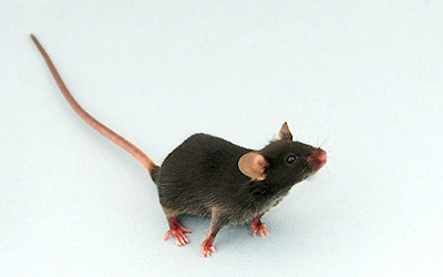
Treatment of mice (Model 11520) with Isoproterenol (β-adrenergic receptor agonist) shows CNS response.
- Multiple transgenic mouse lines were generated through pronuclear microinjection of the CRE-Luc transgene.
- The expression pattern in the founders varied between lineages due to the random integration of the transgene.
- The final CRE-Luc lines were selected to contain peak luciferase expression in a pharmaceutically relevant target tissue (i.e. brain, pancreas, lung, spleen, liver).
- These lines have been tested in assays for reference and exploratory profiling with GPCR ligands.

- Dressler H, Economides K, Favara S, Wu NN, Pang Z, and Polites HG. (2014) The CRE luc Bioluminescence Transgenic Mouse Model for Detecting Ligand Activation of GPCRs. Journal of Biomolecular Screening, 2014, 19: 232.
- Download materials presented at the Keystone Symposium on G Protein-Coupled Receptors: Molecular Mechanisms and Novel Functional Insights, Banff, Canada, February 20, 2012. Presentation by Dr. Greg Polites, Sanofi: "The CRE-Luc Reporter Mouse Model: A Transgenic Bioimaging Mouse Model that can Assay Ligand Activation of GPCRs", February 20, 2012. View the accompanying poster.
- Download the CRE-Luc GPCR Reporter Mouse Platform brochure








.jpg)

.jpg)
.jpg)
.jpg)
.jpg)



.jpg)


.jpg)
.jpg)

.jpg)


.jpg)


.jpg)

.jpg)


