Novel Molecular Mechanisms of Colitis in the Mdr1a KO Mouse
Courtney Ferrebee, PhD
Wednesday, June 2nd, 2021
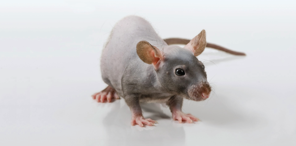
Mdr1a knockout mice are becoming a more frequently utilized colitis model in IBD research due to their translational relevance, microbiome-driven chronic phenotype of colitis, and presence of a functional immune system. However, the primary mechanisms by which colitis appears in the Mdr1a KO mouse are not fully understood. MDR1 is a drug and xenobiotic efflux pump with substrates ranging from steroids to glycolipids1. There are two genetic forms of the protein in the mouse (Abcb1a and 1b or Mdr1a and 1b), while humans (ABCB1 or MDR1) only have one2,3. Expression of Mdr1a is highest in the membrane of absorptive colonocytes, and the presence of the transporter is critical for maintaining intestinal homeostasis through reducing drug absorption and accumulation or elimination in the gut2,4.
Molecular mechanisms of colitis in the Mdr1a Knockout Mouse
Colitis Model
Taconic Health Standard
Mdr1a KO
MPF™
IL-10 KO
EF
Adoptive T Cell Transfer (Rag2 KO on BALB/c background or RAGN12 on B6 background)
EF (Rag2 KO and RAGN12), MPF™ (RAGN12)
Anti-CD40 (Rag2 KO on BALB/c background or RAGN12 on B6 background)
DSS
Dependent on strain of mouse; B6 and BALB/c mice offered in MPF™, EF, and GF
Table 1: Comparison of Taconic health standards in Mdr1a KO mice and other colitis or ID models. To see the accepted microorganisms please visit Taconic's Health Standards.
Mdr1a knockout mice typically develop colitis between 8 to 36 weeks of age. The Mdr1a model presents an interesting story for the molecular mechanisms of colitis compared to other IBD models. Mdr1a mice have a defect in epithelial cell barrier function, which leads to an inability to eliminate the products created by the commensal microbiota (Figure 1)5. Accumulation of these microbial products leads to increased antigen presentation to T cells and considerable hyperactivity. This presents a unique mechanistic phenotype that highlights the importance of targeting the microbiome for therapeutic treatment6. However, this is not the only mechanism of spontaneous colitis in the model. One of the more interesting areas of investigation is the ability of other intestinal metabolites transported by Mdr1a to contribute to the phenotype. Bile acids have been shown to increase intestinal inflammation and modulate Mdr1a function and T effector cells in the intestine7,8. More importantly, the presence of Mdr1a mitigates oxidative stress caused by bile acids on T cells9,10. This presents a colitis mechanism by which loss of Mdr1 prevents intestinal homeostasis by leading to increased oxidative stress, mitochondrial dysfunction, and promotion of inflammation through T cell-mediated colitis11,12. This implicates not only the microbiome but also T cell-driven inflammation and mitochondrial dysfunction as the disease mechanism leading to or worsening epithelial barrier function in Mdr1a KO mice9,13.
Similar to other models, the colitis phenotype is also further aggravated by the presence of pathogenic infection, such as with Helicobacter1,4,14. Therefore, the health profile at an animal facility becomes an important factor in the development of colitis in these mice (Table 1)4. Other environmental insults, such as Dextran sodium sulfate and piroxicam, have also been utilized to induce spontaneous colitis in these mice15. Similar to the Il10 knockout model of IBD, Mdr1a knockouts on different inbred strain backgrounds have varying colitis susceptibility. In Mdr1a knockouts, this may be associated with strain-specific differences in the expression of cytokines and other inflammation-related genes15. For the Mdr1a KO model, FVB mice are genetically more susceptible to permeability changes in the intestine due to loss or aberrant function of Mdr1a and are therefore the most appropriate strain for these studies15.
Histopathology
Histopathological changes in Mdr1a KO mice include a defective mucus barrier due to loss of goblet cells along with increased inflammation from mucosal thickening16. There are also CD4+ T (and other inflammatory) cell infiltrates and crypt abscesses in the colon16. Intestinal inflammation develops at ~12-16 weeks and the pathology is similar to human disease12,17. Mdr1a KO mice have increased levels of myeloperoxidase (a neutrophil accumulation marker) and pro-inflammatory cytokines IL8, IL12, IFNγ, TNFa, and IL1b4,15. Animals present with dysregulated epithelial cell proliferation and leukocyte infiltration of the lamina propria in the large intestine12,17.
Translational relevance and utility
The Mdr1a KO mouse presents with high construct, face, and predictive validity for the study of human IBD and the development of therapeutics. Polymorphisms in MDR1 are strongly associated with ulcerative colitis in humans, and loss of expression has been implicated in the development of the disease3. Barrier dysfunction is very common in human IBD patients, and defective antimicrobial mechanisms and loss of microbial sensing (through TLR2) have a role in Crohn's disease and human ulcerative colitis18-20. Mdr1a KO mice also serve as a platform for investigating cytokine networks (e.g., IL2, IL12, IL17, IL23) and T cell (CD4+, Tregs) involvement in IBD21. Mdr1a mice treated with IL17 neutralizing antibodies have exacerbated colitis while mice treated with IL23 neutralizing antibodies have an attenuated phenotype22. Similar to human clinical outcomes and treatment of ulcerative colitis, studies in Mdr1a mice showed effective treatment of colitis with both anti-IL12 and IL-23 blockades4.
Summary
The Mdr1a KO mouse model is a very useful tool for preclinical IBD research which has been applied across basic and applied studies. This model has high translational utility due to the parallel of molecular mechanisms of the disease (barrier dysfunction, microbiome involvement, and inflammation) to those in humans and the clinical relevance of therapeutic efficacy in this model. Further investigation into Mdr1a's evolutionary role in the colon and as a transporter should be determined in this model. Additional investigation should also explore the expression of Mdr1b in the colon and its potential role in the colitis phenotype.
1. Cario, E. P-glycoprotein multidrug transporter in inflammatory bowel diseases: More questions than answers. World J Gastroentero23, 1513-1520 (2017).
2. Cui, Y. J., Cheng, X., Weaver, Y. M. & Klaassen, C. D. Tissue Distribution, Gender-Divergent Expression, Ontogeny, and Chemical Induction of Multidrug Resistance Transporter Genes (Mdr1a, Mdr1b, Mdr2) in Mice. Drug Metab Dispos37, 203-210 (2009).
3. Brinar, M. et al. MDR1polymorphisms are associated with inflammatory bowel disease in a cohort of Croatian IBD patients. Bmc Gastroenterol13, 57 (2013).
4. Bilsborough, J., Fiorino, M. F. & Henkle, B. W. Select animal models of colitis and their value in predicting clinical efficacy of biological therapies in ulcerative colitis. Expert Opin Drug Dis 1-11 (2020) doi:10.1080/17460441.2021.1851185.
5. Resta-Lenert, S., Smitham, J. & Barrett, K. E. Epithelial dysfunction associated with the development of colitis in conventionally housed mdr1a-/- mice. Am J Physiol-gastr L289, G153-G162 (2005).
6. Berinstein, J., Steiner, C., Higgins, P. The IBD Therapeutic Pipeline is Primed to Produce. Practical Gastro. Inflammatory Bowel Disease, A Practical Approach, 106 (2019).
7. Cao, W. et al. The Xenobiotic Transporter Mdr1 Enforces T Cell Homeostasis in the Presence of Intestinal Bile Acids. Immunity47, 1182-1196.e10 (2017).
8. Sinha, S. R. et al. Dysbiosis-Induced Secondary Bile Acid Deficiency Promotes Intestinal Inflammation. Cell Host Microbe27, 659-670.e5 (2020).
9. Ho, G.-T. et al. MDR1 deficiency impairs mitochondrial homeostasis and promotes intestinal inflammation. Mucosal Immunol11, 120-130 (2018).
10. Izcue, A. & Pabst, O. Mdr1 Saves T Cells from Bile. Immunity47, 1016-1018 (2017).
11. Chen, M. L. et al. Physiological expression and function of the MDR1 transporter in cytotoxic T lymphocytes. J Exp Med217, (2020).
12. Tanner, S. M., Staley, E. M. & Lorenz, R. G. Altered generation of induced regulatory T cells in the FVB.mdr1a-/- mouse model of colitis. Mucosal Immunol6, 309-323 (2013).
13. Rath, E., Moschetta, A. & Haller, D. Mitochondrial function -- gatekeeper of intestinal epithelial cell homeostasis. Nat Rev Gastroentero 15, 497-516 (2018).
14. Maggio-Price, L. et al. Helicobacter bilis Infection Accelerates and H. hepaticus Infection Delays the Development of Colitis in Multiple Drug Resistance-Deficient (mdr1a-/-) Mice. Am J Pathology160, 739-751 (2002).
15. Staley, E. M., Schoeb, T. R. & Lorenz, R. G. Inflamm Bowel Dis15, 684-696 (2009).
16. Panwala, C. M., Jones, J. C. & Viney, J. L. A novel model of inflammatory bowel disease: mice deficient for the multiple drug resistance gene, mdr1a, spontaneously develop colitis. J Immunol Baltim Md 1950161, 5733-44 (1998).
17. Wilk, J. N., Bilsborough, J. & Viney, J. L. The mdr1a-/- mouse model of spontaneous colitis. Immunol Res31, 151-159 (2005).
18. Nalle, S. C. & Turner, J. R. Intestinal barrier loss as a critical pathogenic link between inflammatory bowel disease and graft-versus-host disease. Mucosal Immunol8, 720-730 (2015).
19. Sartor, R. B. Microbial Influences in Inflammatory Bowel Diseases. Gastroenterology134, 577-594 (2008).
20. Ey, B. et al. Loss of TLR2 Worsens Spontaneous Colitis in MDR1A Deficiency through Commensally Induced Pyroptosis. J Immunol190, 5676-5688 (2013).
21. Chen, M. L. & Sundrud, M. S. Cytokine Networks and T-Cell Subsets in Inflammatory Bowel Diseases. Inflamm Bowel Dis22, 1157-1167 (2016).
22. Maxwell, J. R. et al. Differential Roles for Interleukin-23 and Interleukin-17 in Intestinal Immunoregulation. Immunity43, 739-750 (2015).
We’re Here to Help
Experience & Expertise You Can Trust
Taconic Biosciences' model generation team has produced about 5,000 models in the last 15 years, developing a globally-recognized reputation for advancing the work of in vivo researchers. Our scientific program managers are here to help you navigate the complexities of model generation.